Although current guidelines for invasive melanoma without nodal metastases recommend surgery with wide margin excision (wme), use of mms for this disease has increased as well, particularly in early stages. Mohs micrographic surgery for nonmelanoma skin cancers.

Mohs micrographic surgery (mms) is very successful in the treatment of nonmelanoma skin cancer.
Mohs micrographic surgery for melanoma. Evaluating the lateral margins of melanomas using frozen tissue sections is complicated. Mamelak discusses this process with each. Mohs micrographic surgery, frequently shortened to just mohs surgery, was developed in the 1930s by dr.
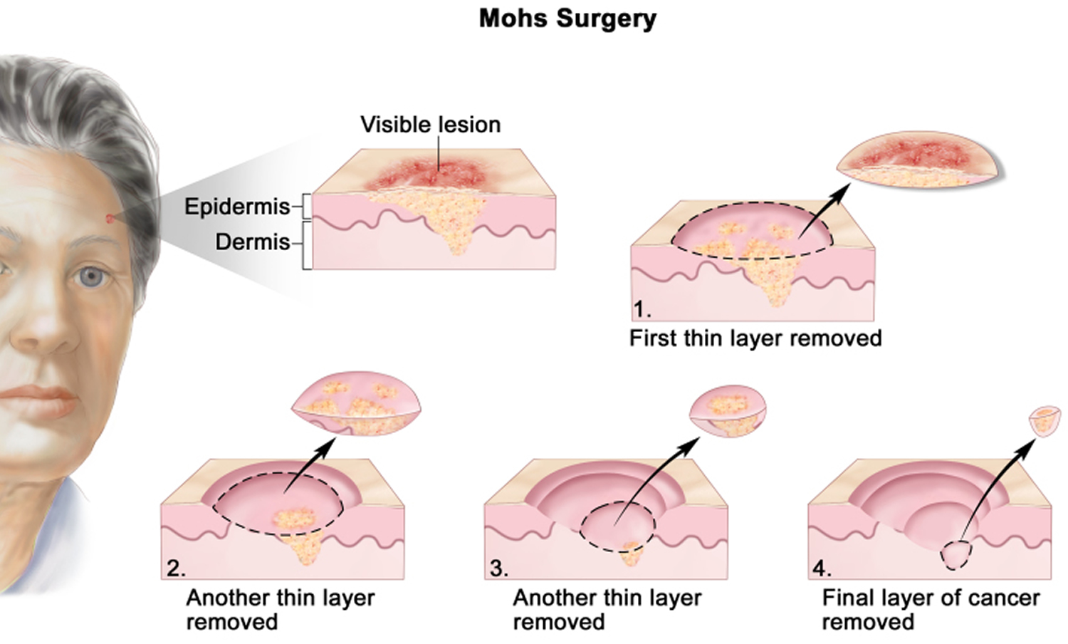
Mohs micrographic surgery is also called margin controlled excision. It offers the highest cure rates while maximizing preservation of healthy tissue. It’s called slow because the patient must wait longer for the results.
Mohs surgery for melanoma insitu: Mohs micrographic surgery (mms) is most useful for: It is done as an outpatient surgery under local anesthesia by dermatology surgeons.
Nelson br, railan d, cohen s. The use of mms for melanoma treatment has yet to become widely accepted owing to difficulties in histologic interpretation, among other factors. El tal ak, abrou ae, stiff ma, mehregan da.
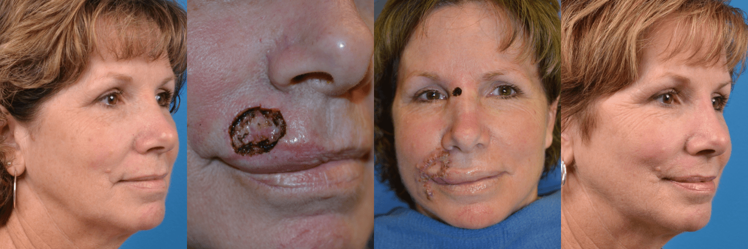
Once your skin cancer has been effectively removed during mohs surgery, you may need a reconstructive procedure, which can typically be performed right after the initial surgery. The principles behind it were developed by dr frederic mohs in the 1930s. Mohs micrographic surgery for nonmelanoma skin cancers.
Examining 100% of the margin using mms improves cure rates. It is a specialist type of surgery that aims to remove all the skin cancer and leave as much healthy skin tissue as possible. The stanford skin cancer program doctors are leaders in.
The use of mms for the treatment of malignant melanoma. Current perspectives on mohs micrographic surgery for melanoma. [ 1] in numerous publications.
When do you have it. Mohs micrographic surgery was associated with improved survival compared with traditional excision in early invasive melanoma, a retrospective study indicated. This method has obvious appeal in treating melanoma.
Mohs micrographic surgery is a highly specialized and precise procedure performed solely by surgeons with advanced training who are both surgeon and pathologist. This type of melanoma stays close to the surface of the skin for a while. Mohs is only used to treat an early melanoma, and it must be a type of melanoma called lentigo malignant melanoma.
Mohs micrographic surgery is primarily used to treat basal and squamous cell carcinomas, but can be used to treat less common tumors including melanoma. Mohs surgery is appropriate when: Few mohs laboratories have the special stains and expertise needed to perform the mohs micrographic technique for melanoma.
Initially, mohs believed that the in situ tissue fixation with zinc Although current guidelines for invasive melanoma without nodal metastases recommend surgery with wide margin excision (wme), use of mms for this disease has increased as well, particularly in early stages. Mohs micrographic surgery (mms), a specialized surgical excision technique used primarily in the treatment of skin cancers, is tissue sparing and provides optimal margin control through evaluation of 100% of both the peripheral and deep margin.
Mohs micrographic surgery, or mohs surgery, is a precise surgical technique in which the complete excision of skin cancer is checked by microscopic margin control. If you have had a more complex mohs surgery, however, your surgeon may need an extra day or more to effectively plan for your reconstruction. The cancer is in an area where it is important to preserve healthy tissue for maximum functional and cosmetic result, such as eyelids, nose, ears, lips, fingers, toes, genitals;
Cherpelis bs, glass lf, ladd s, fenske na. Mohs micrographic surgery (mms) is very successful in the treatment of nonmelanoma skin cancer. Mohs micrographic surgery (mms) has been extensively used and studied for the treatment of nonmelanoma skin cancer, particularly at sites where tissue conservation is vital.
Etzkorn jr, sobanko jf, elenitsas r, et al. Frederic mohs, professor of surgery at the university of. The mohs and dermatologic surgery clinic at stanford is a nationally recognized leader in skin cancer management, mohs micrographic surgery and other dermatologic procedures.
Mohs micrographic surgery for melanoma. Mohs micrographic surgery (mms), a specialized surgical excision technique used primarily in the treatment of skin cancers, is tissue sparing and provides optimal margin control through evaluation of 100% of both the peripheral and deep margin. About mohs surgery for melanoma.
Why choose premier dermatology and mohs surgery atlanta for mohs on melanoma? Mohs surgery is an advanced surgical technique that precisely removes many forms of skin cancer while preserving healthy surrounding tissue. Tissue processing methodology to optimize pathologic staging and margin assessment.
In many cases, this surgery is done at your first appointment at stanford, following a skin biopsy to get a. When treating melanoma, the surgeon uses a modified type of mohs surgery called slow mohs. How is mohs surgery performed?
Mohs micrographic surgery reaches and removes the roots of nonmelanoma and melanoma skin cancers. Since 2012, mohs micrographic surgery (mms) has gained popularity in the treatment of melanoma in situ. Immunostaining in mohs micrographic surgery:
It is commonplace for a mohs surgeon to send a melanoma specimen to an outside laboratory for examination of the margins, causing the. We investigated the potential benefits of slow mohs micrographic surgery (mms) for acral melanomas. As it is a specialised type of surgery, your doctor might refer you to another hospital to have the treatment.
Mohs micrographic surgery for melanoma: